
In the standard ResNet's case the classes are the 1000 different objects that it is trained to recognize. Classifications(classes) are the different objects the neural network is trained to recognize. First, the Linear Layer maps the features processed by the other layers to the classifications given. There are two notable layers in this network: linear layer and softmax layer. In addition, I set the program to output the top 10 probabilities of what it thought the image was. It correctly predicts that the image is a golden retriever. The immense computing power, time resources, and knowledge dedicated to perfect this network made it a great neural network to use for transfer learning.īelow is an example of the ResNet 50 in action. I used ResNet 50, an architecture released in 2015 by Microsoft Research Asia this network was originally trained on 1.2 million images with 1000 classes. Improve generalization in another setting"-(, Deep Learning, What has been learned in one setting is exploited to

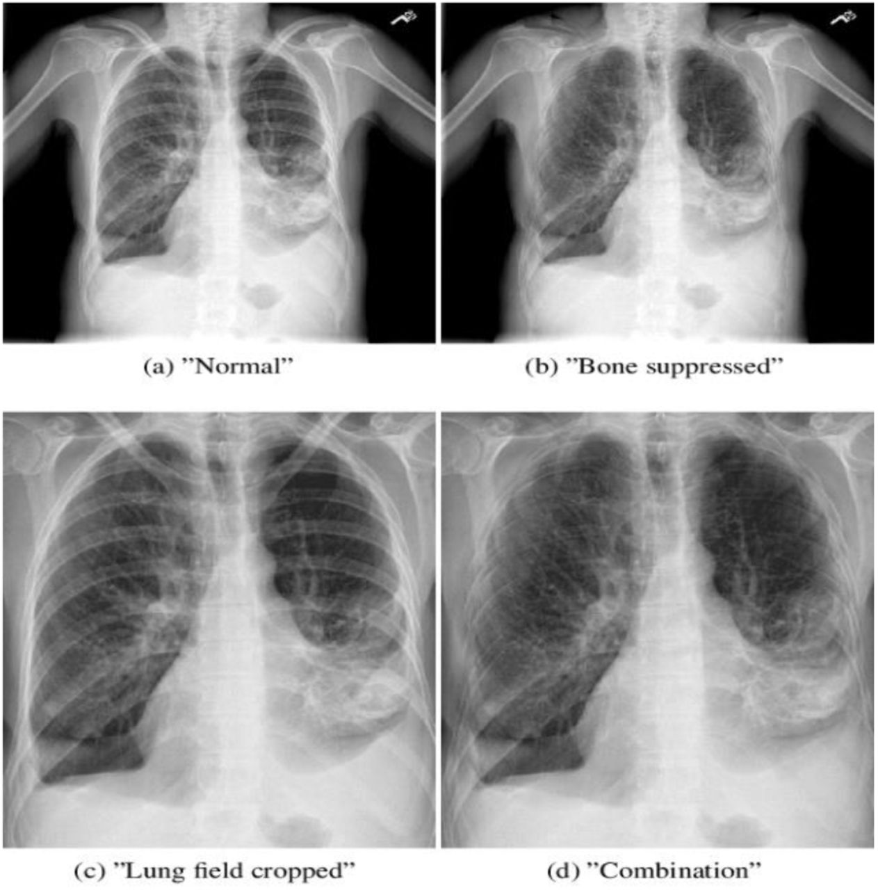
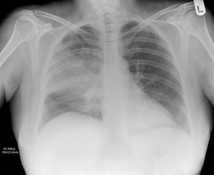
"Transfer learning and domain adaptation refer to the situation where Transfer learning is the process of using a pre-trained model as a starting point as a model for a second task. I used transfer learning to create my neural network. Chest X-rays of pneumonia patients are characterized by loss of diaphramatic shadows, and evidence of pulmonary consolidation(an area of the lung that has filled with liquid instead of air). Thanks to Paul Timothy Mooney for making the dataset available on Kaggle.īelow is an example of an infiltrate present in a chest X-ray.īelow is an example of how chest X-rays of the three classifications generally look. Thank you to Daniel Kermany, Daniel Zhang, and Michael Goldbaum for creating and labeling the dataset. My project uses a convolutional neural network to diagnose the type of pneumonia that a patient has and. A diagnosis is determined by infiltrates, or white spots, present in a chest X-ray. Since pneumonia, is an airspace disease, patients' air spaces are filled with bacteria, viruses, pus, and other microorganisms. Chest X-rays have long been reliable diagnostic tools for pneumonia. Dutch researchers, who published their findings in the European Respiratory Journal, found that of 140 patients who had their pneumonia diagnosed by x-ray, doctors initially thought only 41 of them had the severe lung infection. Conventional diagnosis methods like examining patient medical history and symptoms have their shortcomings. Early and proper diagnosis can tremendously decrease mortality rate.

Although the severity of pneumonia can vary, young children, seniors, and people with a weakened immune system are the most vulnerable.The CDC reports that of the close to 540,000 cases of pneumonia each year, 50,000 people die. Bacterial pneumonia, the more common type, is caused by bacteria that multiplies in the lungs Viral Pneumonia is caused by an array of viruses, the most common one being Influenza. Pneumonia is an infection that causes inflammation in one or both of the lungs it is induced by a variety of organisms: bacteria, viruses, and fungus.


 0 kommentar(er)
